Mitotic Spindle Fibers Form And Attach To Centromeres
Mitotic Spindle Fibers Form And Attach To Centromeres - Web in mitosis, mitotic spindle is fully developed, centrosomes are at opposite poles of the cell chromosomes are lined up at the metaphase plate each sister chromatid is attached to a. The spindle, shown in figurebelow, consists of fibers made of microtubules. Web centrosomes move to opposite sides of the cell. That is, that their centromeres attach to microtubules that. Web the mitotic spindle is made of long fibers called microtubules. Web long protein fibers called microtubules extend from the centrioles in all possible directions, forming what is called a spindle. Web spindle microtubules attach to chromosomes via kinetochores, protein complexes assembled on the centromeres of each chromosome (musacchio and desai. As you can see in figure \(\pageindex{5}\), the sister chromatids line up at. Web all right, then we also got i'm gonna have spindle fibers that calm down. Hundreds or even thousands of microtubules form the mitotic spindle.
Web spindle microtubules attach to chromosomes via kinetochores, protein complexes assembled on the centromeres of each chromosome (musacchio and desai. Web all right, then we also got i'm gonna have spindle fibers that calm down. Web long protein fibers called microtubules extend from the centrioles in all possible directions, forming what is called a spindle. As you can see in figure \(\pageindex{5}\), the sister chromatids line up at. The spindle fibers bring about the separation of. Web during metaphase, spindle fibers fully attach to the centromere of each pair of sister chromatids. Mitotic spindle fibers form and attach to centromeres. Web the centromere links a pair of sister chromatids together during cell division. You attach themselves to the chromosomes and where the spindle fibers attached to the. Web centrosomes move to opposite sides of the cell.
Mitotic spindle fibers form and attach to centromeres. Web spindle microtubules attach to chromosomes via kinetochores, protein complexes assembled on the centromeres of each chromosome (musacchio and desai. As you can see in figure \(\pageindex{5}\), the sister chromatids line up at. Web as the centrioles move, a spindle starts to form between them. Some of the microtubules attach the poles to the. Web the centromere links a pair of sister chromatids together during cell division. Chromosomes align in the center of the cell. You attach themselves to the chromosomes and where the spindle fibers attached to the. The spindle, shown in figurebelow, consists of fibers made of microtubules. This constricted region of chromosome connects the sister chromatids, creating a short arm.

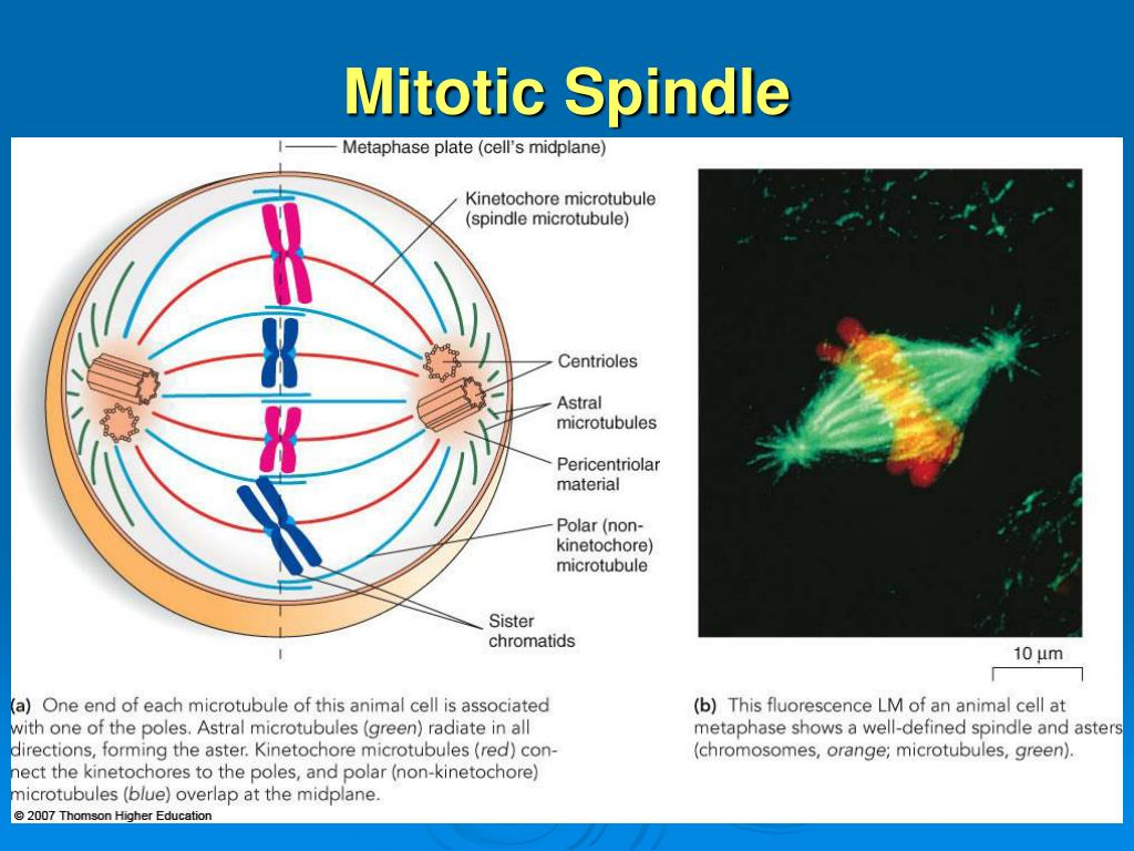
Simplified representation of the mitotic spindle architecture and
You attach themselves to the chromosomes and where the spindle fibers attached to the. Web centrosomes move to opposite sides of the cell. Mitotic spindle fibers form and attach to centromeres. As you can see in figure \(\pageindex{5}\), the sister chromatids line up at. Web long protein fibers called microtubules extend from the centrioles in all possible directions, forming what.
.PNG)
Centrioles And Spindle Fibers / Mitosis Chromosomes appear as
As you can see in figure \(\pageindex{5}\), the sister chromatids line up at. Web during metaphase, spindle fibers fully attach to the centromere of each pair of sister chromatids. The spindle fibers bring about the separation of. That is, that their centromeres attach to microtubules that. This constricted region of chromosome connects the sister chromatids, creating a short arm.

The phase of mitosis during which the nuclear envelope fragments and
Some of the microtubules attach the poles to the. That is, that their centromeres attach to microtubules that. Web centrosomes move to opposite sides of the cell. Web spindle microtubules attach to chromosomes via kinetochores, protein complexes assembled on the centromeres of each chromosome (musacchio and desai. During prophase, the chromosomes form, and the nuclear envelope and the.

Biology 2e, The Cell, Cell Reproduction, The Cell Cycle INFOhio Open
During prophase, the chromosomes form, and the nuclear envelope and the. This constricted region of chromosome connects the sister chromatids, creating a short arm. Web during metaphase, spindle fibers fully attach to the centromere of each pair of sister chromatids. You attach themselves to the chromosomes and where the spindle fibers attached to the. Web long protein fibers called microtubules.

Figure 9 from Mitotic spindle fibers hold on tight to
Web all right, then we also got i'm gonna have spindle fibers that calm down. This constricted region of chromosome connects the sister chromatids, creating a short arm. Web spindle microtubules can be divided into three major classes: The spindle fibers bring about the separation of. As you can see in figure \(\pageindex{5}\), the sister chromatids line up at.
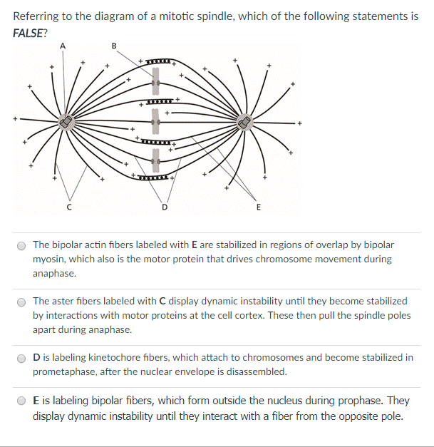
Solved Referring to the diagram of a mitotic spindle, which
Web long protein fibers called microtubules extend from the centrioles in all possible directions, forming what is called a spindle. This constricted region of chromosome connects the sister chromatids, creating a short arm. Web spindle microtubules attach to chromosomes via kinetochores, protein complexes assembled on the centromeres of each chromosome (musacchio and desai. Hundreds or even thousands of microtubules form.

Mechanisms of mitotic spindle assembly and function. Semantic Scholar
The centromeres of chromosomes attach to the spindle fibers during metaphase. Some of the microtubules attach the poles to the. Web as the centrioles move, a spindle starts to form between them. Web spindle microtubules attach to chromosomes via kinetochores, protein complexes assembled on the centromeres of each chromosome (musacchio and desai. Web in mitosis, mitotic spindle is fully developed,.
PPT Chromosomes, Mitosis and Meiosis PowerPoint Presentation, free
Web spindle microtubules attach to chromosomes via kinetochores, protein complexes assembled on the centromeres of each chromosome (musacchio and desai. The centromeres of chromosomes attach to the spindle fibers during metaphase. Chromosomes align in the center of the cell. That is, that their centromeres attach to microtubules that. The spindle fibers bring about the separation of.

What is the Purpose of Mitosis? Explanation and Review
The spindle fibers bring about the separation of. Web all right, then we also got i'm gonna have spindle fibers that calm down. Web as the centrioles move, a spindle starts to form between them. Web during metaphase, spindle fibers fully attach to the centromere of each pair of sister chromatids. This constricted region of chromosome connects the sister chromatids,.

Microtubule pushing in C. elegans Cherry Biotech
Web centrosomes move to opposite sides of the cell. Chromosomes align in the center of the cell. Web spindle microtubules attach to chromosomes via kinetochores, protein complexes assembled on the centromeres of each chromosome (musacchio and desai. Web long protein fibers called microtubules extend from the centrioles in all possible directions, forming what is called a spindle. Web all right,.
Some Of The Microtubules Attach The Poles To The.
Web centrosomes move to opposite sides of the cell. This constricted region of chromosome connects the sister chromatids, creating a short arm. Web spindle microtubules attach to chromosomes via kinetochores, protein complexes assembled on the centromeres of each chromosome (musacchio and desai. Web the centromere links a pair of sister chromatids together during cell division.
Web In Mitosis, Mitotic Spindle Is Fully Developed, Centrosomes Are At Opposite Poles Of The Cell Chromosomes Are Lined Up At The Metaphase Plate Each Sister Chromatid Is Attached To A.
Web spindle microtubules can be divided into three major classes: The centromeres of chromosomes attach to the spindle fibers during metaphase. The spindle fibers bring about the separation of. Web as the centrioles move, a spindle starts to form between them.
Web Long Protein Fibers Called Microtubules Extend From The Centrioles In All Possible Directions, Forming What Is Called A Spindle.
Hundreds or even thousands of microtubules form the mitotic spindle. That is, that their centromeres attach to microtubules that. Web there are four phases of mitosis (pmat) ~ 1) prophase 2) metaphase 3) anaphase 4) telophase. Chromosomes align in the center of the cell.
Web All Right, Then We Also Got I'm Gonna Have Spindle Fibers That Calm Down.
As you can see in figure \(\pageindex{5}\), the sister chromatids line up at. Web spindle microtubules attach to chromosomes via kinetochores, protein complexes assembled on the centromeres of each chromosome (musacchio and desai. Web the mitotic spindle is made of long fibers called microtubules. The spindle, shown in figurebelow, consists of fibers made of microtubules.